Published On Nov 19, 2019
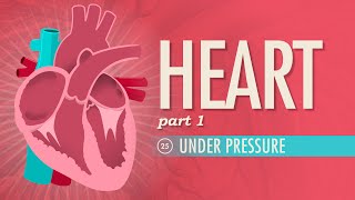
The heart is a vital organ, which is the size of your fist,
It’s crowned by the great vessels, of four chambers does consist,
Cardiac muscle does pump, connective tissue supports,
Valves separate the chambers, vessels direct transport.
It’s behind the ribs and sternum, in between the lungs,
In front of the spinal column, with vertebrae like rungs,
On top of the diaphragm, and slightly to the left,
In the cardiac impression, which makes room for its heft!
The central compartment of the thoracic cavity,
The mediastinum has important anatomy,
Know it is surrounded by loose connective tissue,
Fixed to the pericardium, holding the heart in situ!
The heart’s top is called the base, the bottom the apex,
Oh why did anatomists decide to perplex?
The pericardium lubricates and prevents from infection,
The fibrous and serous layers, provide this protection!
The serous layers enclose, the pericardial fluid,
For reducing heartbeat friction, it is well reputed!
The fibrous pericardium is connective tissue, OK?
Attached to the diaphragm, sternum and vertebrae!
Serous pericardium, layers parietal and visceral!
The visceral one on the heart’s surface is visible!
The myocardium is the muscular middle layer,
For pumping blood this section is the main player!
The coronary vessels go from the outside in,
Supplying the myocardium, which is found within,
The fibrous cardiac skeleton is made of connective tissue,
Specifically collagen to support muscle tissue!
The endocardium, the innermost of the layers,
Made up of endothelium, it lines the heart and chambers!
Veins bring blood toward the heart, the arteries away,
It’s not about oxygenation, so don’t be led astray!
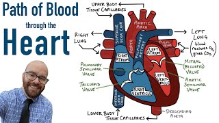
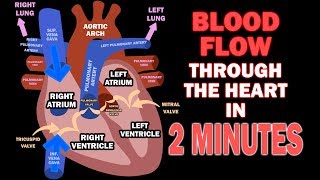
The two vena cavas bring blood to our hearts,
To enter the right atrium, and so blood’s journey starts!
Blood from the coronary sinus, from the myocardium drains,
Collecting all the blood from coronary vessel domains!
The atrioventricular valves cause separation,
Between the atria and ventricles is their location!
The tricuspid valve has three cusps, the mitral one has two,
They open up when they need to let blood pass through!
These cusps have strings attached, the chordae tendineae,
Tethered to papillary muscle, which keeps blood at bay!
For when the heart contracts, the tendineae are taut,
Closing off the atria so return the blood cannot!
So only through the next valve can the blood now flow!
The pulmonary valve, which has three cusps to show!
It leads to the pulmonary artery, which carries blood to the lungs,
Returning to the heart, with oxygen it comes!
The pulmonary veins bring blood back to the heart,
So oxygenated blood to the body does depart!
But first the blood must enter the left ventricle,
Through a valve with just two cusps, called the mitral!
So pulmonary circulation comes to an end,
From heart to lungs and back, on that you can depend!
Now the blood goes through systemic circulation!
Man, this organ never gets any relaxation!
The left ventricle and myocardium is bigger,
Which makes sense, as it requires more vigor,
Because it pumps to the body at large,
While the lungs are the left ventricle’s only charge!
The aortic valve, door to the largest artery,
It has three cusps leading to the aorta as you see,
The auricles of the atria, seen from outside,
Are muscular pouches, that more space for blood provide!
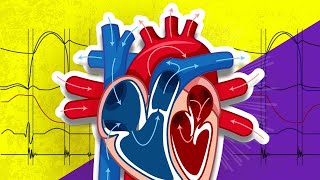
The sinoatrial node is the heart’s pacemaker,
The atrioventricular node is its helpful neighbour!
The SA node triggers the atria to contract!
The AV node makes the ventricles pump once packed!
Music/Lyrics/Visual © Neural Academy
Heart model by https://www.turbosquid.com/3d-models/...