Published On Jun 17, 2019
Structure of the Eye in a Snap! Unlock the full A-level Biology course at http://bit.ly/2K4JC0y created by Adam Tildesley, Biology expert at SnapRevise and graduate of Cambridge University.
SnapRevise is the UK’s leading A-level and GCSE revision & exam preparation resource offering comprehensive video courses created by A* Oxbridge tutors. Our courses are designed around the OCR, AQA, SNAB, Edexcel B, WJEC, CIE and IAL exam boards, concisely covering all the important concepts required by each specification. In addition to all the content videos, our courses include hundreds of exam question videos, where we show you how to tackle questions and walk you through step by step how to score full marks.
Sign up today and together, let’s make A-level Biology a walk in the park!
The key points covered of this video include:
1. Protecting the Eye
2. Bending Light
3. Getting to the Retina
4. Location of Photoreceptors
Protecting the Eye
The eye is an important and delicate sensory organ, and needs to be protected. The conjunctiva is a layer of cells on the outside of the eye. It protects the cornea, which is a transparent layer of cells. The rest of the eye is protected by a hard layer called sclera.
Bending Light
Light coming from a single point spreads out. If the retina received light that was spread out the image would be unfocused. To focus the image, the eye bends the light coming in. Most of the bending is done by the cornea, which bends light by a fixed amount. The rest is achieved by the lens. The lens can bend light different amounts, depending on how far away we’re trying to see. This is achieved by increasing or decreasing the thickness of the lens. This movement is carried out by the ciliary muscles.
Outer Structure of the Eye
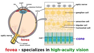
The eye can also control how much light enters by adjusting the pupil size. This is done by muscles in the iris. Once light gets through the pupil, it travels through a transparent jelly called the vitreous humour. Finally the light is able to reach the retina, which absorbs and detects the light. Behind the retina is an absorbent layer called the choroid. This stops light being reflected behind the retina. This means the only light being absorbed by the retina is light coming from outside the eye, instead of reflected from behind the retina.
Location of Photoreceptors
The retina contains photoreceptors, which are receptor cells that detect light. The region with most photoreceptors is called the fovea. There are no photoreceptors in the blind spot. This is because this contains the optic nerve, where neurones carry signals out of the eye to the brain.
Summary
The eye is protected by the sclera and the conjunctiva
Light is bent by the cornea and the retina
This allows the image to be focused
Pupil size is controlled by the iris muscles
Light travels through the vitreous humour to the retina, which contains the photoreceptors
Signals are taken out of the eye to the brain via the optic nerve
Behind the retina is the choroid which prevents any light being reflected back