Published On Oct 16, 2017
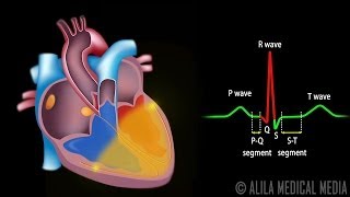
(USMLE topics, cardiology) Phases of the cardiac cycle. The Wiggers diagram explained.
Purchase PDF here: https://www.alilamedicalmedia.com/-/g...
Purchase a license to download a non-watermarked copy of this video here: https://www.alilamedicalmedia.com/-/g...
©Alila Medical Media. All rights reserved.
Voice by: Sue Stern.
Support us on Patreon and get FREE downloads and other great rewards: patreon.com/AlilaMedicalMedia
The cycle is initiated with the firing of the SA node that stimulates the atria to depolarize. This is represented by the P-wave on the ECG. Atrial contraction starts shortly after the P-wave begins, and causes the pressure within the atria to increase, FORCING blood into the ventricles. Atrial contraction, however, only accounts for a FRACTION of ventricular filling, because at this point, the ventricles are ALREADY almost full due to PASSIVE blood flow DOWN the ventricles through the OPEN AV valves.
As atrial contraction completes, atrial pressure begins to FALL, REVERSING the pressure gradient across the AV valves, causing them to CLOSE. The closing of the AV valves produces the first heart sound, S1, and marks the beginning of SYSTOLE. At this point, ventricular depolarization, represented by the QRS complex, is half way through, and the ventricles start to contract, RAPIDLY building UP pressures inside the ventricles. For a moment, however, the semilunar valves remain closed, and the ventricles contract within a CLOSED space. This phase is referred to as isovolumetric contraction, because NO blood is ejected and ventricular volume is UN-changed.
Ventricular ejection starts when ventricular pressures EXCEED the pressures within the aorta and pulmonary artery; the aortic and pulmonic valves OPEN and blood is EJECTED out of the ventricles. This is the RAPID ejection phase.
As ventricular repolarization, reflected by the T-wave, begins, ventricular pressure starts to FALL and the force of ejection is REDUCED.
When ventricular pressures drop BELOW aortic and pulmonary pressures, the semilunar valves CLOSE, marking the end of systole and beginning of diastole. Closure of semilunar valves produces the second heart sound, S2.
The first part of diastole is, again, isovolumetric, as the ventricles relax with ALL valves CLOSED. Ventricular pressure drops RAPIDLY but their volumes remain UNchanged.
Meanwhile, the atria are being filled with blood and atrial pressures RISE slowly. Ventricular FILLING starts when ventricular pressures drop BELOW atrial pressures, causing the AV valve to open, allowing blood to flow DOWN the ventricles PASSIVELY.
All images/videos by Alila Medical Media are for information purposes ONLY and are NOT intended to replace professional medical advice, diagnosis or treatment. Always seek the advice of a qualified healthcare provider with any questions you may have regarding a medical condition.