Published On Jan 24, 2017
(USMLE topics, cardiology) Cardiac action potential in pacemaker cells and contractile myocytes, electrophysiology of a heartbeat.
Purchase PDF (script of this video +images) here: https://www.alilamedicalmedia.com/-/g...
Purchase a license to download a non-watermarked copy of this video here: part 1 (pacemaker): https://www.alilamedicalmedia.com/-/g...
part 2 (myocytes): https://www.alilamedicalmedia.com/-/g...
©Alila Medical Media. All rights reserved.
Voice by: Sue Stern.
Support us on Patreon and get FREE downloads and other great rewards: / posts
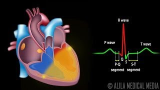
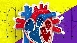
The heart is essentially a muscle that contracts and pumps blood. It consists of specialized muscle cells called cardiac myocytes. The contraction of these cells is initiated by electrical impulses, known as action potentials. The impulses start from a small group of myocytes called the PACEMAKER cells, which constitute the cardiac conduction system. The cells of the SA node fire SPONTANEOUSLY, generating action potentials that spread though the contractile myocytes of the atria. The myocytes are connected by gap junctions. This enables electrical coupling of neighboring cells. Pacemaker cells and contractile myocytes exhibit different forms of action potentials.
The pacemaker cells of the SA node SPONTANEOUSLY fire about 80 action potentials per minute, each of which sets off a heartbeat. Pacemaker cells do NOT have a TRUE RESTING potential. The voltage starts at about -60mV and SPONTANEOUSLY moves upward until it reaches the threshold of -40mV. This is due to action of so-called “FUNNY” currents present ONLY in pacemaker cells. Funny channels open when membrane voltage becomes lower than -40mV and allow slow influx of sodium. The resulting DE-polarization is known as “pacemaker potential”. Calcium channels open, calcium ions flow into the cell further DE-polarizing the membrane. This results in the rising phase. At peak, potassium channels open, calcium channels inactivate, potassium ions leave the cell and the voltage returns to -60mV. This is falling phase of the action potential.
Contractile myocytes have a different set of ion channels. Their sarcoplasmic reticulum, the SR, stores a large amount of calcium. They also contain myofibrils. The contractile cells have a stable resting potential of -90mV and depolarize ONLY when stimulated. When a cell is DE-polarized, positive ions leak through the gap junctions to the adjacent cell and bring the membrane voltage of this cell up to the threshold of -70mV. FAST sodium channels open creating a rapid sodium influx and a sharp rise in voltage. This is the depolarizing phase. L-type, or SLOW, calcium channels also open at -40mV, causing a slow but steady influx. Sodium channels close quickly, voltage-gated potassium channels open and these result in a small decrease in membrane potential, known as EARLY RE-polarization phase. The calcium channels remain open and the potassium efflux is eventually balanced by the calcium influx. This keeps the membrane potential relatively stable for about 200 msec resulting in the PLATEAU phase, characteristic of cardiac action potentials. Calcium is crucial in coupling electrical excitation to physical muscle contraction. The influx of calcium from the extracellular fluid triggers a MUCH greater calcium release from the SR, in a process known as “calcium-induced calcium release". Calcium sets off muscle contraction by “sliding filament mechanism”. Calcium channels close, potassium efflux predominates and membrane voltage returns to its resting value. The absolute refractory period is much longer in cardiac muscle. This is essential in preventing summation and tetanus.
All images/videos by Alila Medical Media are for information purposes ONLY and are NOT intended to replace professional medical advice, diagnosis or treatment. Always seek the advice of a qualified healthcare provider with any questions you may have regarding a medical condition.